Understanding tooth decay is essential for maintaining oral health and preventing serious dental problems. This article, inspired by the comprehensive explanation from Osmosis from Elsevier, delves into the causes, symptoms, diagnosis, pathology, and treatment of tooth decay and cavities. Whether you are a healthcare professional or simply interested in dental health, this guide offers a clear and detailed overview of this common condition.
Table of Contents
- What is Tooth Decay?
- Anatomy of the Tooth and Surrounding Structures
- The Battle Between Demineralization and Remineralization
- How Acidity Leads to Tooth Decay
- Progression of Tooth Decay
- Symptoms and Diagnosis
- Prevention and Treatment
- Summary
- Frequently Asked Questions (FAQ)
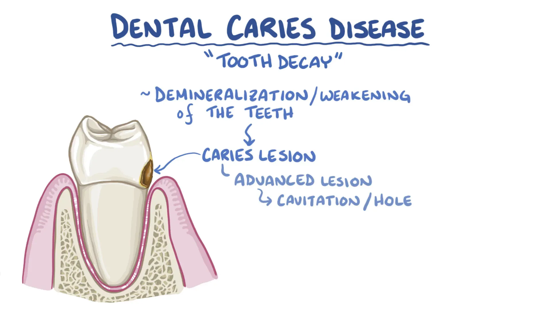
What is Tooth Decay?
Tooth decay, also known as dental caries, is the process of demineralization or weakening of the teeth. The end result of untreated tooth decay is the formation of a caries lesion, commonly referred to as a cavity. Advanced decay leads to the formation of a visible cavity or hole on the tooth surface, indicating the breakdown of the tooth structure.
Anatomy of the Tooth and Surrounding Structures
To fully understand tooth decay, it's important to start with the tooth's anatomy and its supporting structures. In the mouth:
- The bone beneath the lower teeth is the mandible (lower jaw), and the bone above the upper teeth is the maxilla (upper jaw).
- Each tooth sits in a socket called the alveolus, which is lined internally by the periodontal ligament that supports the tooth.
- Externally, the alveolus is covered by soft supporting tissue known as the gingiva (gums), which also covers the root surface up to the cemento-enamel junction, where the enamel meets the cementum.
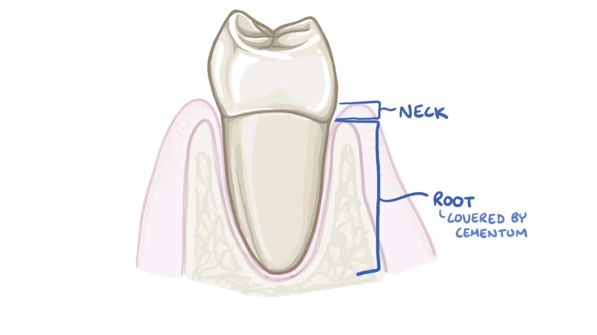
The tooth itself is divided into parts:
- Root: Embedded in the alveolus and covered by cementum, a bone-like material to which periodontal ligament fibers attach.
- Neck: The transition area between the root and the crown.
- Crown: The visible part of the tooth above the gum line, covered by enamel, which is the hardest tissue in the human body due to its high mineral content.
Enamel is produced by specialized cells called ameloblasts during tooth development, but once the tooth erupts into the mouth, these cells die, meaning enamel cannot be regenerated naturally.
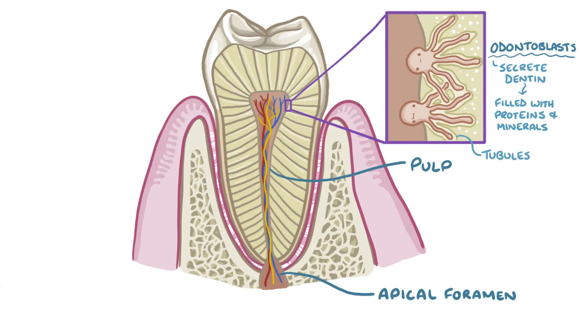
Internal Tooth Structure
Inside the tooth, blood vessels and nerves enter through a small opening at the tip of the root called the apical foramen. These extend into the soft center of the tooth called the pulp, which provides nourishment and sensory function.
Surrounding the pulp is dentin, a mineralized tissue secreted by odontoblasts. These cells have long processes extending into tiny tubules within the dentin, allowing sensory nerves to transmit signals from the pulp to the enamel-dentin junction.
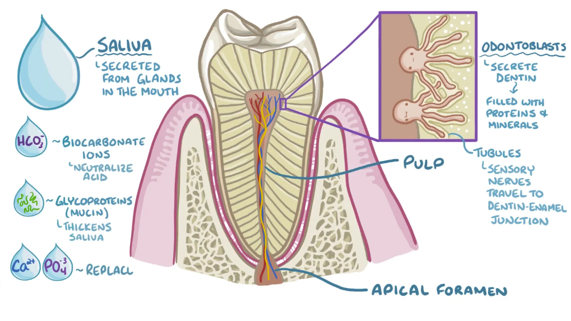
The Role of Saliva
Saliva, secreted by glands in the mouth, plays a critical protective role. It contains bicarbonate ions that neutralize acids, mucin proteins that give saliva its thickness, and minerals like calcium and phosphate that help remineralize tooth surfaces.
The Battle Between Demineralization and Remineralization
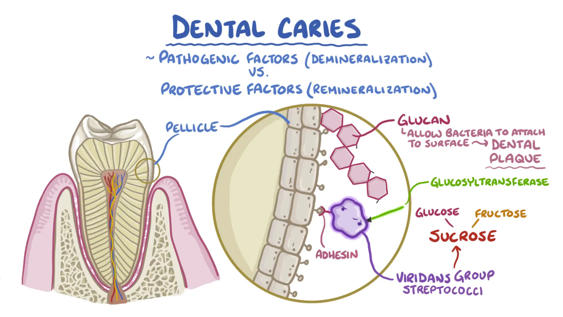
Tooth decay is essentially a tug-of-war between forces that remove minerals from the tooth (demineralization) and those that restore them (remineralization). A protective layer called the pellicle covers the teeth, providing a surface for early bacterial colonizers, such as streptococcus sanguinis, to adhere.
These bacteria use surface proteins called adhesins to stick to the pellicle. When exposed to dietary sugar, particularly sucrose (table sugar), bacteria produce an enzyme called glucosyltransferase that splits sucrose into glucose and fructose. Fructose is metabolized for energy, while glucose molecules are linked into long chains called glucans, which help bacteria firmly attach to the tooth and form a sticky biofilm known as dental plaque.
This biofilm is a complex community where bacteria communicate chemically and cooperate, creating a structured environment with specialized functions—much like an ant colony with tunnels and chambers.
How Acidity Leads to Tooth Decay
The key factor tipping the balance towards decay is a drop in pH around the tooth surface. When the pH falls below about 5.5, enamel begins to lose minerals through demineralization. Conditions that reduce saliva production or its buffering capacity—such as Sjögren’s syndrome, radiation therapy, acid reflux, or certain medications—can promote this acidic environment.
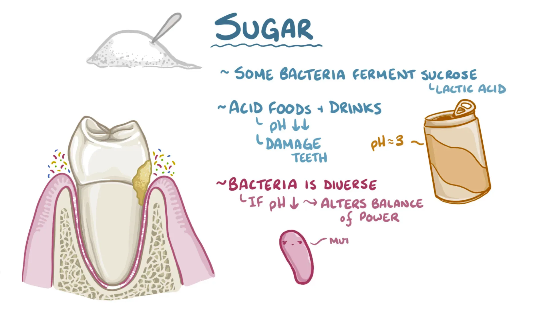
Sugars are fermented by acid-producing bacteria like mutans streptococci and lactobacilli, generating lactic acid. Acidic foods and drinks further lower the pH. For example, soda typically has a pH around 3, which harms teeth both by direct acid erosion and by feeding acid-producing bacteria.
As plaque thickens and oxygen levels decrease near the enamel surface, anaerobic bacteria thrive and continue producing acid even without oxygen, rapidly lowering the pH below the critical threshold and causing mineral loss from enamel crystals.
Progression of Tooth Decay
Repeated acid attacks dissolve calcium and phosphate ions from enamel, thinning it until it collapses, similar to walking on thin ice. Healthy enamel’s microscopic pores are too small for bacteria to penetrate, but once enamel is cavitated, bacteria can invade the underlying dentin.
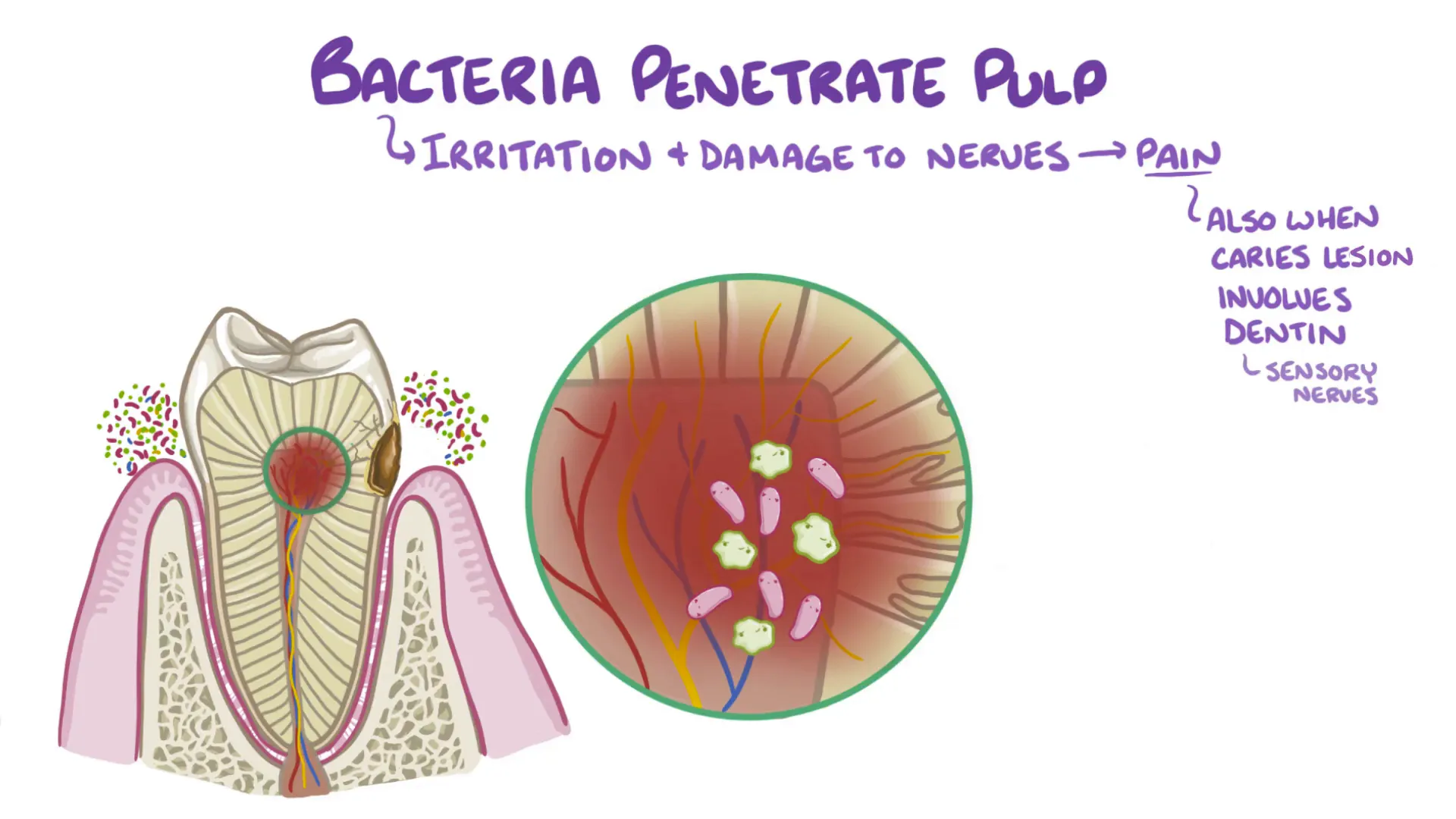
Decay in dentin, known as softened dentin, usually requires restoration with fillings. If untreated, decay progresses toward the pulp, causing inflammation and nerve irritation, often resulting in pain. Eventually, bacteria enter the root canal system, filling the inner tooth and potentially leading to abscess formation.
Decay can also begin below the gum line when gums recede or plaque accumulates near the root, where cementum and dentin are less mineralized and more vulnerable. Demineralization in these areas occurs at higher pH levels (between 6.2 and 6.8).
Symptoms and Diagnosis
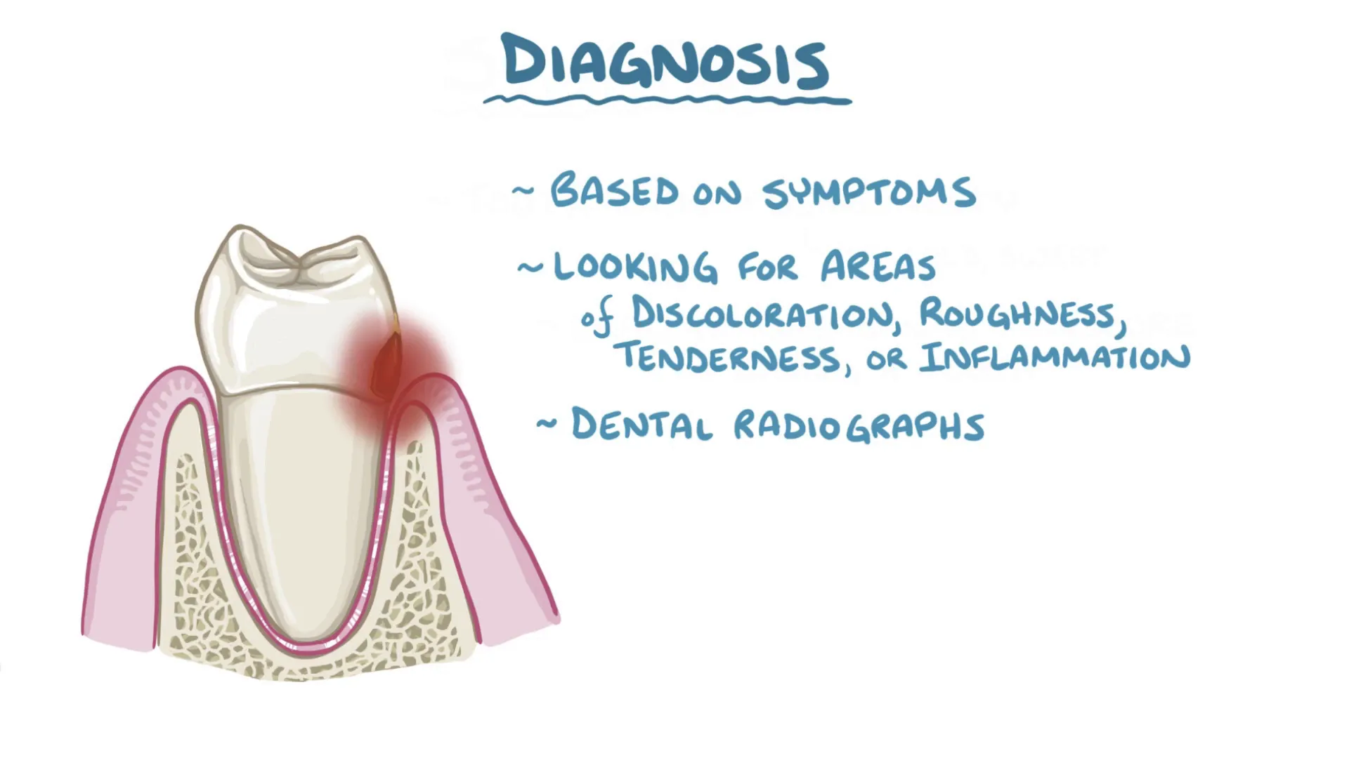
Symptoms of tooth decay include:
- Tooth pain or sensitivity, especially when chewing or consuming hot, cold, or sweet foods and drinks.
- Visible discoloration or roughness on tooth surfaces.
- Inflammation or discomfort in the gums.
Diagnosis typically involves clinical examination looking for signs of decay and the use of dental X-rays to detect hidden lesions.
Prevention and Treatment
Preventing tooth decay starts with assessing individual risk factors, such as a history of cavities, saliva production issues, presence of acidogenic bacteria, and dietary habits like frequent snacking or high sugar intake.
Treatment focuses on:
- Reducing risk factors: Using antimicrobial mouthwashes to reduce harmful bacteria and increasing protective factors like saliva substitutes.
- Mechanical removal of plaque: Brushing and flossing physically remove biofilm buildup.
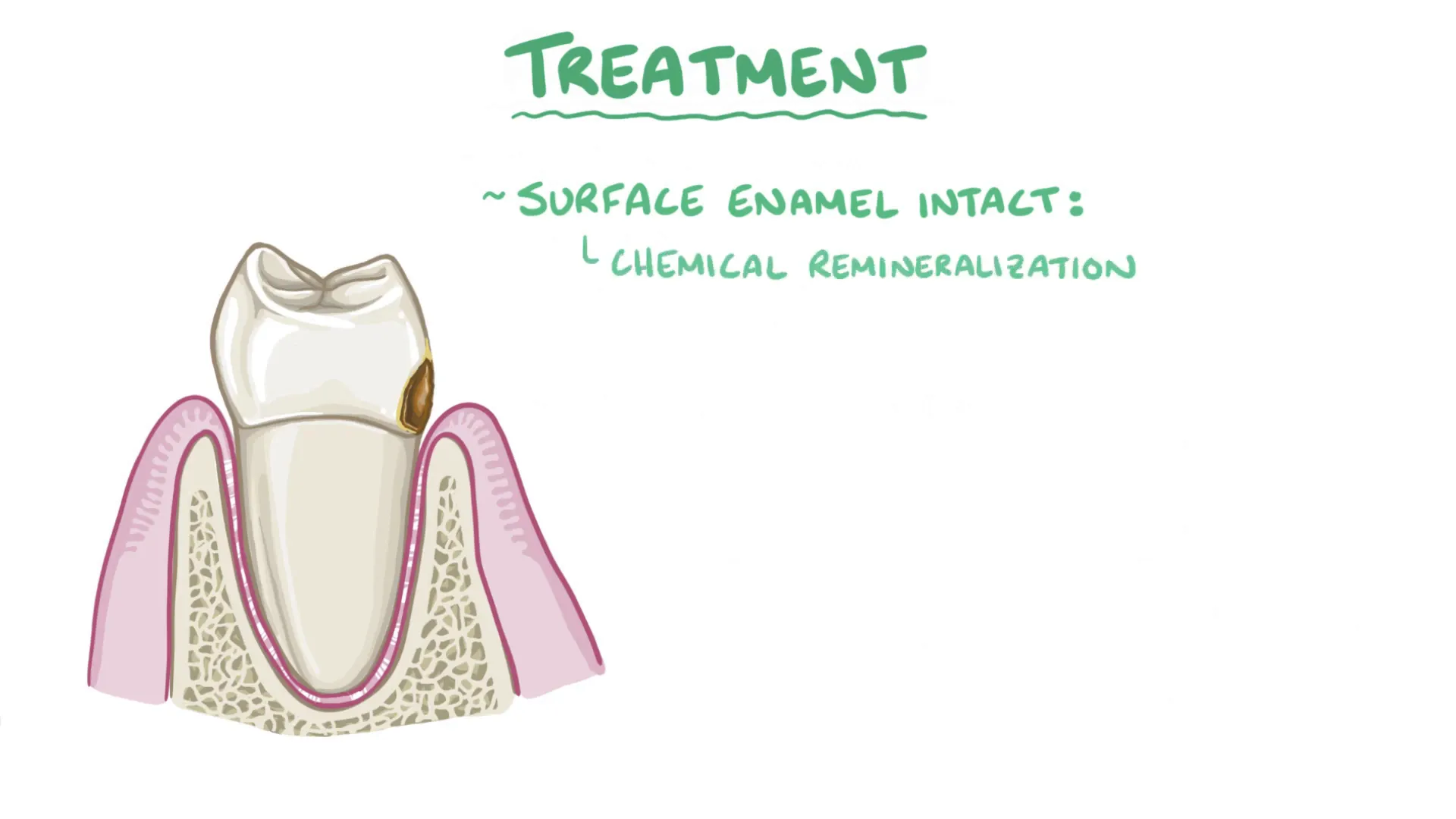
- Fluoride application: Fluoride promotes remineralization and can reverse early enamel lesions.
If the enamel surface remains intact, chemical remineralization can restore the tooth without drilling. However, once a cavity penetrates enamel into dentin, the decayed tissue must be removed and replaced with a filling to restore function.
In severe cases where decay extensively damages the tooth, a dental crown may be necessary to protect the remaining structure.
While restorative treatments are effective, they do not address the underlying causes of decay, so ongoing prevention is essential to avoid new cavities.
Summary
Tooth decay results from a complex interplay between dietary sugars, acid-producing bacteria, and the tooth's mineral balance. When the oral environment becomes acidic (pH below 5.5 for enamel), minerals dissolve, leading to cavities. Bacterial biofilms (dental plaque) play a crucial role in this process.
Diagnosis involves clinical and radiographic examination, and treatment ranges from preventive remineralization to restorative procedures like fillings and crowns. Prevention through good oral hygiene, diet management, and regular dental care remains the cornerstone of maintaining healthy teeth.
Frequently Asked Questions (FAQ)
What causes tooth decay?
Tooth decay is caused primarily by acid-producing bacteria in dental plaque, which ferment sugars from the diet and lower the pH around the teeth, leading to mineral loss from enamel and dentin.
Can tooth enamel repair itself?
Enamel cannot regenerate once formed because the cells that create it die after tooth eruption. However, early enamel lesions can be repaired through remineralization using saliva and fluoride.
How does saliva protect teeth?
Saliva neutralizes acids with bicarbonate ions, provides minerals like calcium and phosphate for remineralization, and helps wash away food particles and bacteria.
What are common symptoms of tooth decay?
Symptoms include tooth sensitivity or pain, especially when eating or drinking hot, cold, or sweet substances, visible discoloration, and sometimes gum inflammation.
How is tooth decay treated?
Treatment depends on severity and includes fluoride applications, antimicrobial rinses, fillings for cavities, and crowns for extensive damage. Prevention through good oral hygiene is vital.
Can diet affect tooth decay risk?
Yes, frequent consumption of sugary and acidic foods and drinks increases the risk by feeding acidogenic bacteria and lowering oral pH.
Is it possible to prevent tooth decay entirely?
While complete prevention is challenging, maintaining good oral hygiene, reducing sugar intake, regular dental visits, and using fluoride can significantly reduce the risk.